Bone Cross Section Diagram / Cross Section Of Bone Full Size Png Download Seekpng - This is a cross section through decalcified bone.
byAdmin•
0
Bone Cross Section Diagram / Cross Section Of Bone Full Size Png Download Seekpng - This is a cross section through decalcified bone.. A cross section of a human long bone. For example, to read this diagram literally, since the cartilage can be seen inside the cutaway section of bone, it incorrectly indicates that the cartilage in fact goes through the bone structure, rather than just being found around the bone end. Related posts of cross section of human bone diagram bone in arm pictures. This diagram depicts anatomy of the human eye cross section view.human anatomy diagrams show internal organs, cells, systems, conditions, symptoms and sickness information and/or tips for healthy living. Bone test anatomy and physiology 12 photos of the bone test anatomy and physiology anatomy and physiology bone lab test, anatomy and physiology bone markings test, anatomy and physiology bone practical test, anatomy and physiology bone tissue test, anatomy and physiology test on bone tissue, bone, anatomy and.
.and hyaline cartilage slides, b) describe the differences you observed between the elastic cartilage and hyaline cartilage slides palaglapu styles data table 3. It is the most complete reference of human anatomy available on web, ipad, iphone and android devices. Bone in arm pictures 12 photos of the bone in arm pictures bone cancer arm pictures, pictures of bone cancer in arm, bone, bone cancer arm pictures, pictures of bone cancer in arm. That's here these 2 diagrams come into play. Free online quiz compact bone microscope slide labeled.
9 1 Bone Structure And Function Medicine Libretexts from med.libretexts.org Cross section of long bone diagram | quizlet. (b) in this micrograph of the osteon, you can clearly see the concentric lamellae and central canals. Explore over 6700 anatomic structures and more than 670 000 translated medical labels. Smartdraw includes 1000s of professional healthcare and anatomy chart templates that you can modify and make your own. Shop bone cross section diagram label created by chartsanddiagrams. Related posts of cross section of human bone diagram bone in arm pictures. New users enjoy 60% off. It seems confusing and misleading.
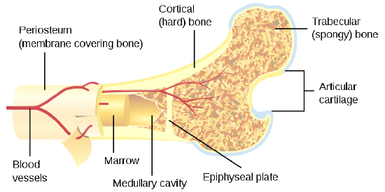
There are trabeculae in spongy bone which gives its sponge like appearance.
Personalize it with photos & text or purchase as is! I don't like way you've shown the cartilage. The bladder, like the stomach, is an expandable sac that contracts with inner folds when it is empty. Bone in arm pictures 12 photos of the bone in arm pictures bone cancer arm pictures, pictures of bone cancer in arm, bone, bone cancer arm pictures, pictures of bone cancer in arm. It seems confusing and misleading. Smartdraw includes 1000s of professional healthcare and anatomy chart templates that you can modify and make your own. This is a cross section through decalcified bone. Free online quiz compact bone microscope slide labeled. Cross section of long bone diagram | quizlet. (b) in this micrograph of the osteon, you can clearly see the concentric lamellae and central canals. .and hyaline cartilage slides, b) describe the differences you observed between the elastic cartilage and hyaline cartilage slides palaglapu styles data table 3. Bone test anatomy and physiology 12 photos of the bone test anatomy and physiology anatomy and physiology bone lab test, anatomy and physiology bone markings test, anatomy and physiology bone practical test, anatomy and physiology bone tissue test, anatomy and physiology test on bone tissue, bone, anatomy and. Smear) 6 envelopes containing color images of epithelial 2.
(b) in this micrograph of the osteon, you can clearly see the concentric lamellae and central canals. (b) in this micrograph of the osteon, you can clearly see the concentric lamellae and central canals. Cross section of a long bone. Bone test anatomy and physiology 12 photos of the bone test anatomy and physiology anatomy and physiology bone lab test, anatomy and physiology bone markings test, anatomy and physiology bone practical test, anatomy and physiology bone tissue test, anatomy and physiology test on bone tissue, bone, anatomy and. It is the most complete reference of human anatomy available on web, ipad, iphone and android devices.
6 3 Bone Structure Anatomy Physiology from open.oregonstate.education It seems confusing and misleading. (b) in this micrograph of the osteon, you can see the concentric lamellae around the central canals. Cross section of long bone diagram | quizlet. Smartdraw includes 1000s of professional healthcare and anatomy chart templates that you can modify and make your own. In simple terms, a tree can be described as a bundle of vessels, its walls composed of cellulose glued together with lignin. Explore over 6700 anatomic structures and more than 670 000 translated medical labels. There are trabeculae in spongy bone which gives its sponge like appearance. Bone decalcification is the removal of the mineral component using an acid, leaving the bone soft and easy to cut.
The star of the show (brain) is easily recognizable because it appears highly convoluted, full of ridges (gyri) and indentations (sulci).the paired thalami appear as two circular masses in the midline, forming the walls of the third ventricle.the neurocranium appears as a meshwork (trabecular.
Diagram orienting yourself within such a cross section is easy. Human bone, cross section diagram of femur showing osteon, veins, marrow. The star of the show (brain) is easily recognizable because it appears highly convoluted, full of ridges (gyri) and indentations (sulci).the paired thalami appear as two circular masses in the midline, forming the walls of the third ventricle.the neurocranium appears as a meshwork (trabecular. Wood is a porous three dimensional, hydroscopic, interconnecting matrix of cellulose, hemicelluloses and lignin. Bone marrow is the soft, highly vascular and flexible connective tissue within bone cavities which serve as the primary site of new blood cell production or bone marrow is the primary source of pluripotent stem cells that give rise to all hemopoietic cells (blood cells) including lymphocytes. Smartdraw includes 1000s of professional healthcare and anatomy chart templates that you can modify and make your own. The bladder, like the stomach, is an expandable sac that contracts with inner folds when it is empty. These vessels or cells transport food and waste products through the tree. (b) in this micrograph of the osteon, you can clearly see the concentric lamellae and central canals. Ct, mri, radiographs, anatomic diagrams and nuclear images. Smear) 6 envelopes containing color images of epithelial 2. Cross section through the thalamus: I don't like way you've shown the cartilage.
The inner lining of the bladder tucks into the folds and expand out to. Browse 4,294 bone cross section stock photos and images available, or search for human bone cross section to find more great stock photos and pictures. It is the most complete reference of human anatomy available on web, ipad, iphone and android devices. Diagram orienting yourself within such a cross section is easy. Smear) 6 envelopes containing color images of epithelial 2.
Unit 4 Skeletal System General Human Anatomy And Physiology from sites.google.com It is the most complete reference of human anatomy available on web, ipad, iphone and android devices. Smartdraw includes 1000s of professional healthcare and anatomy chart templates that you can modify and make your own. Personalize it with photos & text or purchase as is! Cross section through the thalamus: Shop bone cross section diagram label created by chartsanddiagrams. This diagram depicts anatomy of the human eye cross section view.human anatomy diagrams show internal organs, cells, systems, conditions, symptoms and sickness information and/or tips for healthy living. Diagram with articular cartilage, marrow, medullary cavity and periosteum. The first illustrates the anatomy of a window and frame.
Bone marrow is the soft, highly vascular and flexible connective tissue within bone cavities which serve as the primary site of new blood cell production or bone marrow is the primary source of pluripotent stem cells that give rise to all hemopoietic cells (blood cells) including lymphocytes.
Smear) 6 envelopes containing color images of epithelial 2. Explore over 6700 anatomic structures and more than 670 000 translated medical labels. Personalize it with photos & text or purchase as is! That's here these 2 diagrams come into play. Diagram with articular cartilage, marrow, medullary cavity and periosteum. .and hyaline cartilage slides, b) describe the differences you observed between the elastic cartilage and hyaline cartilage slides palaglapu styles data table 3. There are trabeculae in spongy bone which gives its sponge like appearance. Jump to navigation jump to search. Free online quiz compact bone microscope slide labeled. The bladder, like the stomach, is an expandable sac that contracts with inner folds when it is empty. Cross section of a long bone. Cross section of long bone diagram | quizlet. Shop bone cross section diagram label created by chartsanddiagrams.
Compact bone and spongy bone: bone cross section. Wood is a porous three dimensional, hydroscopic, interconnecting matrix of cellulose, hemicelluloses and lignin.